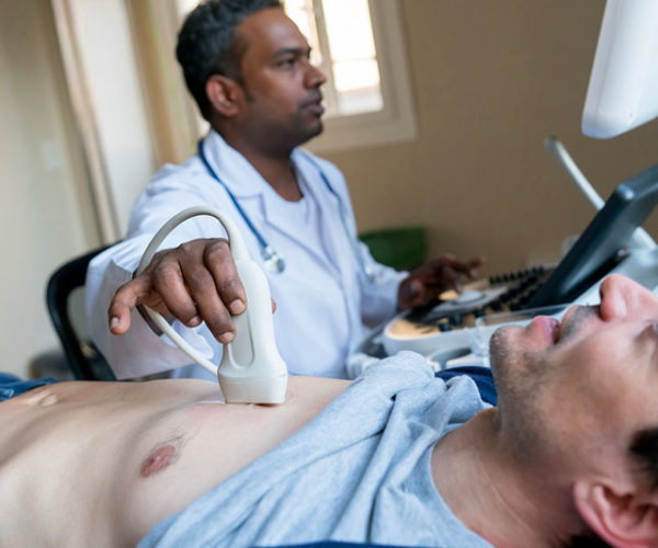
A significant cardiac murmur may prompt a clinician to order echocardiography in a patient with chest discomfort or breathing difficulties. This device creates pictures of your heart using sound waves. This routine procedure gives doctors a visual of your heart as it pumps. If your doctor suspects cardiac trouble, they may utilize an echocardiogram Upper East Side pictures as a starting point in their diagnosis. Echocardiograms come in various forms, and the one you get will depend on your doctor’s specific requirements. All echocardiograms pose little, if any, danger to the patient.
Among the numerous advantages of having echocardiography performed are:
There are little to no risks
In contrast to diagnostic techniques that employ radiation to obtain data, echocardiograms pose no danger to the patient. Because it is a non-invasive test, you won’t have to worry about an actual cut’s discomfort and potential problems.
Though transthoracic echocardiography may cause some pain, it is entirely safe. It is possible to have a painful throat after transesophageal echocardiography, but it should go away in at least a few hours. Stress echocardiography aims to measure the heart’s response to physical exertion. While a higher heart rate is expected during a stress echocardiogram, heart attacks and other serious complications are relatively uncommon.
Monitoring treatment safety and effectiveness
Echocardiography may also track disease progression and response to therapy or surgical outcomes. For instance, specific echocardiography is utilized for follow-up monitoring after coronary artery bypass grafting (CABG). Monitoring heart failure patients routinely using echocardiography is also beneficial. Patients with cancer treatment when cardiotoxicity is possible are also routinely monitored using echocardiograms.
Disease and abnormality screening
Echocardiography will give conclusive evidence if your doctor suspects you have a major cardiac condition. Echocardiograms are used to evaluate heart function, diagnose heart illness, and monitor the development of cardiac masses. It is essential to keep an eye on patients with heart valve problems now classified as mild or minor in case they progress to the moderate or severe stages. A heart attack, stroke, or clot might occur if these problems are neglected. Some of the conditions that an echocardiogram can spot include:
- Heart valve disease: Blood flow irregularities inside the heart due to dysfunction of one or more heart valves. If the valves become constricted, blood cannot pass through the heart or be pumped to the lungs and the rest of the body. In addition, the valves may become defective, allowing blood to escape backward. If you suspect an infection in the tissue that lines your heart’s valves, an echocardiogram is your best bet for a definitive diagnosis.
- Atherosclerosis: Plaque buildup in the arteries due to fats and other chemicals in the circulation. It may disrupt the normal motion of the heart’s walls and affect the heart’s ability to pump blood. An echocardiogram can catch it early enough.
- Aneurysm: Dilation and weakness of the aorta or cardiac muscle. A rupture of the aneurysm may occur.
Your heart is one of the vital organs in your body. You want it to function at its best consistently, and one of the ways of ensuring that is by seeking an echocardiogram. It monitors your heart’s state and warns you if something is amiss from where you can take the relevant action.


